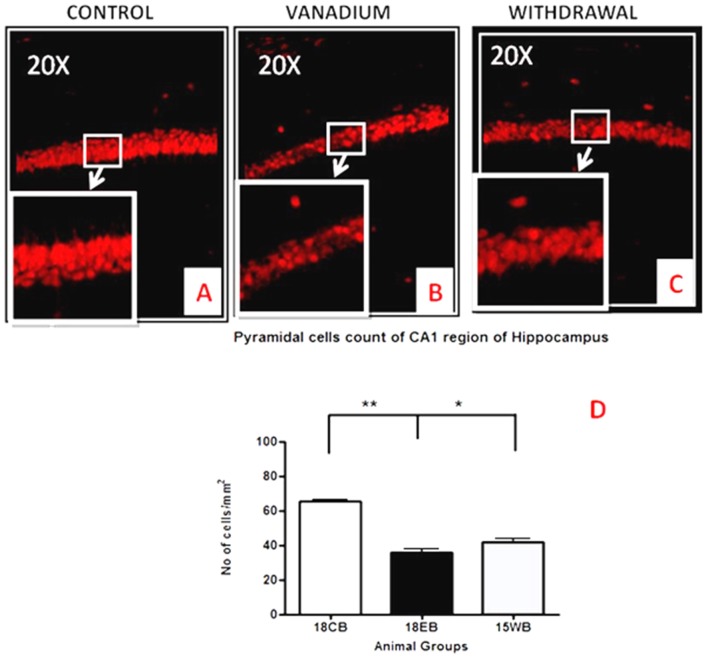
Figure 10.
Pyramidal cell loss of the dorsal hippocampal CA1 region after chronic vanadium exposure. NeuN immuno-histochemistry showed evidence of neuronal loss after 18 months of vanadium treatments. Quantitative analysis (D) revealed significant (**P < 0.001) decrease in the no of pyramidal neurones in the exposed groups (B) relative to the control (A) while the withdrawal groups (C) showed reversal effect with significant (*P < 0.05) increase in neuronal no relative to vanadium exposed groups (B). Magnification: ×200, inset: ×400.

