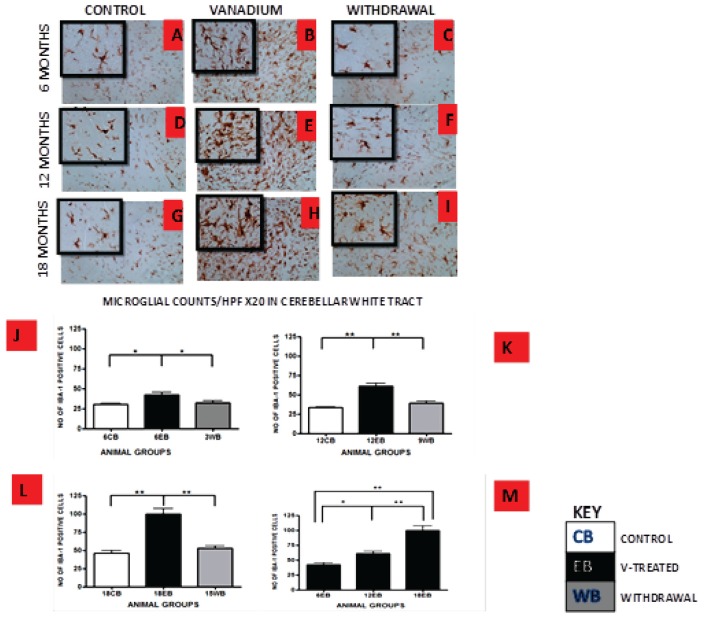
Figure 5.
IBA-1 immuno-stained white matter of cerebellum of vanadium exposed (B,E,H), age matched control (A,D,G) and withdrawal group (C,F,I) mice after intermittent vanadium treatments for 6, 12 and 18 months revealed microglial activation identified by an enlarged cell body with several short, thickened processes, relative to the matched controls with longer, finer branches while the withdrawal groups showed better morphology relative to vanadium exposed groups. (J–L) The number of IBA-1 positive cells in hippocampal CA1 region, and cerebellum were significantly increased in all the vanadium exposed groups compared to controls, while the withdrawal groups showed reversal effect. Persistent microglia activation throughout the exposure increases to phagocytic state indicated by amoeboid isoform. (B,E,H) Microglia activation increases into advanced age with increasing vanadium exposure (M; 6EB vs. 18EB = **p < 0.01), (ANOVA: *P < 0.05, **p < 0.01). Magnification: ×200 inset: ×400.

