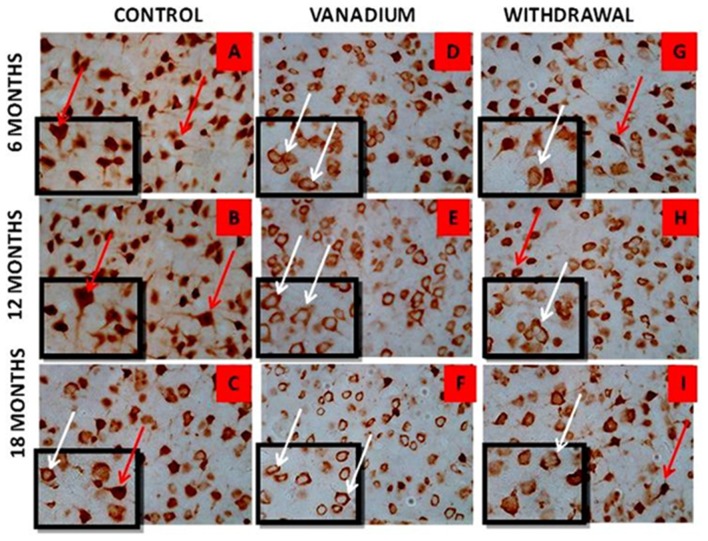
Figure 6.
NeuN immuno-histochemistry revealed the cytotoxic effect of vanadium on the pyramidal cells of the prefrontal cortex after chronic exposure. The cortical pyramidal cells showed morphological alterations including pyknosis, cell clustering, loss of layering pattern and cytoplasmic vacuolation in the vanadium exposed groups (white arrows in B,E,H) relative to the control (red arrows in A,D) with normal neuronal morphology. The withdrawal groups (C,F,I) showed reversal effect with less cellular toxicity relative to vanadium exposed groups. The vanadium cytotoxicity was also observed in the aged control mice brain (white arrows in G), indicative of neuronal degeneration seen also at early vanadium exposure. Magnification: ×400, inset: ×600.

