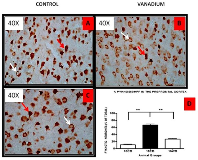
Figure 7.

HPF photomicrographs showing the pyramidal cells of the prefrontal cortex using the NeuN immunohistochemistry. No remarkable abnormality was observed in the cortical sections from animals of the control groups. The normal neurons were identified by their rounded and pale nuclei (see A red arrow), whereas degenerating neurons had smaller cell bodies and pyknotic nuclei (see B white arrow). There was evidence of vacuolation of neuropil surrounding the degenerating neurons. The withdrawal brain (C) showed lesscellular pathology relative to the exposed brains. Quantitative analysis (D) showed that the mean % pyknosis of vanadium exposed groups were significantly (**P < 0.001) elevated relative to the control brain while the withdrawal groups were significantly (**P < 0.001) less than vanadium exposed groups. Magnification: ×400.
