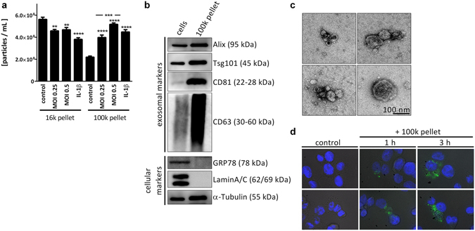
Figure 1.

L. pneumophila infection increases the secretion of EVs in THP-1 cells. (a) Amount of EVs in response to L. pneumophila infection. THP-1 cells were treated with IL-1β (1 ng/mL) or infected with L. pneumophila (MOI 0.25 or 0.5, respectively) for 24 h. NTA was performed with the distinct particle fractions separated by differential centrifugation. (b) Western blot for exosomal marker proteins. Whole cell lysate or 100 k pellet derived from uninfected THP-1 cells were used. Equal protein amounts were loaded. (c) Transmission electron microscopy with 100 k pellet. Purified EVs from THP-1 cells were fixed and visualized after negative staining with uranyl acetate. Scale bar represents 100 nm. (d) Uptake of A549-derived EVs by THP-1 cells. A549 cells were stained with the membrane dye PKH67. EVs were collected (100 k pellet) and incubated with THP-1 cells for 1 or 3 h, respectively. Nuclei were stained by DAPI and pictures were taken with an original magnification of 630x. A: Data are shown as mean + SEM of three independent experiments. **p < 0.01, ***p < 0.001, ****p < 0.0001. B–D: representative results.
