Figure 4.

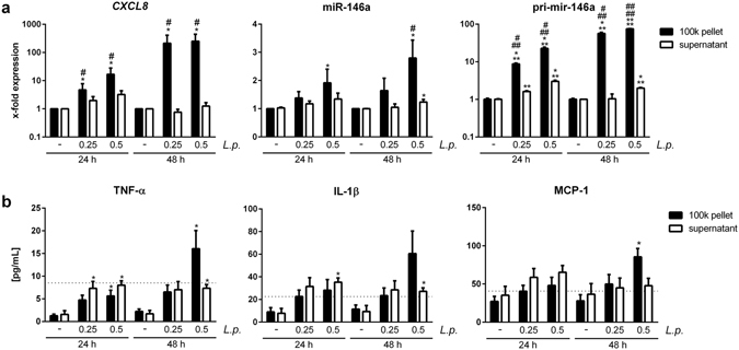
L. pneumophila induced EVs elicit a pro-inflammatory response in non-infected THP-1 cells. (a,b) Response of THP-1 cells to vesicle-free supernatant or 100 k pellet from L. pneumophila-infected THP-1 cells. THP-1 cells were stimulated with vesicle-free supernatant or 100 k pellet from THP-1 cells infected with L. pneumophila (MOI 0.25 and 0.5, respectively) for 24 h. Recipient THP-1 cells were incubated for 24 or 48 h, respectively. (a) qPCR was performed for expression of CXCL8, miR-146a and pri-mir-146a. (b) Magnetic multiplex ELISA was performed. Results for TNF-α, IL-1β and MCP-1 are shown. Dashed lines indicate the input of the cytokine contained in the vesicle-free supernatant of infected THP-1 cells (MOI 0.5) before adding it to the recipient cells. Data are shown as mean + SEM of at least three independent experiments. *p < 0.05. *Compared to corresponding control, # compared to corresponding supernatant treated sample.
