
Figure 7.
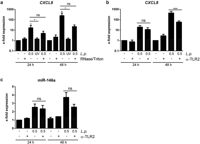
Viable L. pneumophila are critical for response of THP-1 cells to EVs. (a) Response of THP-1 cells to EVs from L. pneumophila-infected THP-1 cells. THP-1 cells were infected with L. pneumophila or stimulated with UV-inactivated L. pneumophila (both MOI 0.5 for 24 h). 100 k pellets were generated and left untreated or digested with RNase A (0.2 µg/µL) in the presence of 0.3% Triton X-100 for 1 h at 37 °C. The prepared EVs were added to THP-1 cells for 24 or 48 h, respectively, exposing the cells to a subcritical final Triton X-100 concentration of 0.003%. Expression of CXCL8 was determined. (b,c) TLR2 dependent response of THP-1 cells to EVs from L. pneumophila-infected THP-1. THP-1 cells were infected with L. pneumophila (MOI 0.5 for 24 h) and the 100 k pellet was generated. Recipient THP-1 cells were pre-incubated for 90 min with a TLR2 blocking antibody (α-TLR2,+) or a control antibody (−; both 20 µg/mL). The EVs were added for 24 or 48 h, respectively. Expression of CXCL8 (b) and miR-146a (c) was determined. Data are shown as mean + SEM of three independent experiments. *p < 0.05, ns: not significant.