
Figure 3.
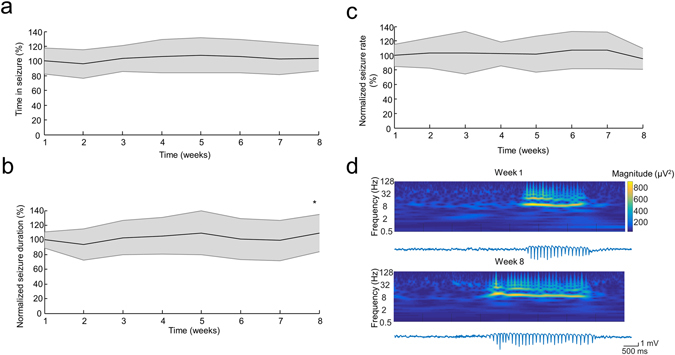
Seizure characteristics and signal quality of the non-treated animals. (a,b and c) Group data shows the Time in Seizure (a), average seizure duration (b) and seizure rate (c) over the weeks of observation (each measure is normalized to its corresponding mean ‘Week 1’ value for each animal). Shaded area represents ± SD. (d) Example LFP traces and continuous wavelet spectra of the recordings of one representative rat demonstrating the stable signal quality over the weeks. *p < 0.01 vs Week 2, one-way ANOVA (a,c), Kruskal-Wallis test (b), both followed by post hoc HSD.