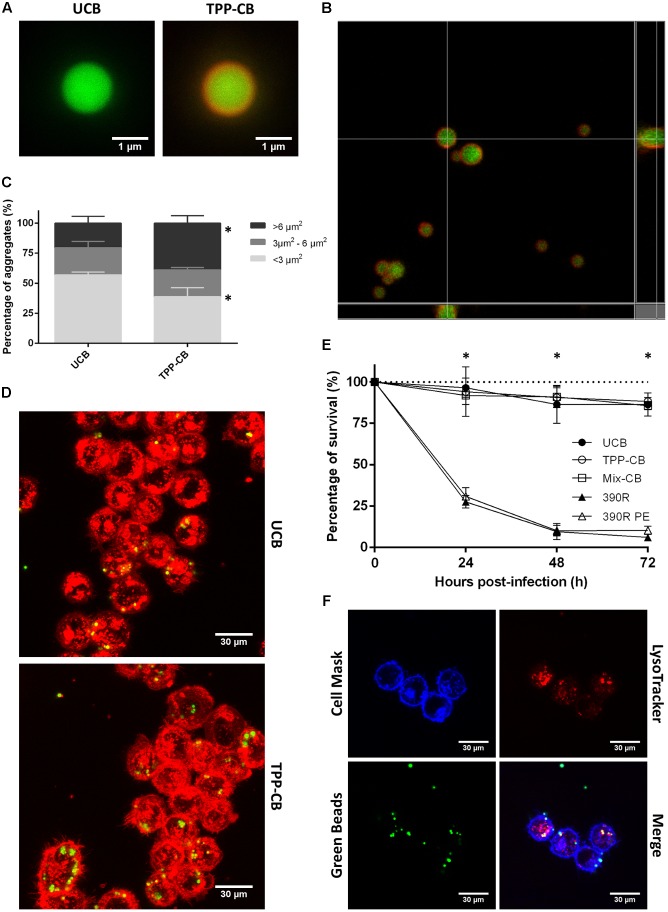
FIGURE 5.
Beads are covered by TPP, and they do not have a toxic effect when in contact with macrophages. (A,B) Beads are in green and TPP-coating is observed as the red covering, owing to staining with Nile Red. Bar size in (A) 1 μm. (C) Results of the study of bead aggregation. ∗p < 0.05 in multiple t-test. The results represent the mean ± SD of triplicate preparations. (D) Example of the images obtained by CLSM that were used to study the aggregation of the beads; macrophages phagocytose both types of beads. Bar size 30 μm. (E) Viability of macrophages treated with uncoated beads; TPP A-coated beads; beads coated with TPP A, compound Y and Z; 390R M. abscessus strain and 390R M. abscessus PE treated. Significant differences were detected only between macrophages treated with beads and macrophages infected with bacteria (∗p < 0.05). The data are representative of one out of three independent experiments. (F) Images of colocalization of beads and acidic vesicles. Macrophages in blue stained with Cell Mask Deep Red, beads in green and acidic vesicles in red with LysoTracker Red. Bar size 30 μm. UCB (Uncoated beads), TPP-CB (TPP A-coated beads), Mix-CB (beads coated with TPP A, compound Y and Z), 390R (M. abscessus 390R stain), 390R PE (M. abscessus 390R treated with PE).

