
Figure 7.
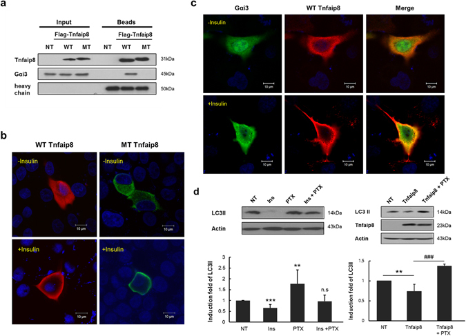
Interaction between wild-type mTnfaip8 and the triple mutant with Gαi3. (a) Co-immunoprecipitation assay using Flag-tagged wild-type mTnfaip8 (WT) and the Flag-tagged mTnfaip8 triple mutant (MT) was performed in Hepa 1-6 cells as described in Materials & Methods. As expected, the mTnfaip8 triple mutant with PE deficiency barely interacted with Gαi3, whereas wild-type mTnfaip8 maintained normal interaction with Gαi3. (b) HepG2 cells were transfected with Flag-mTnfaip8 (WT, red) and Flag-mTnfaip8 triple mutant (MT, green), followed by insulin treatment. Their subcellular relocalization was monitored by confocal microscopy. Representative images are shown here. Both cytoplasmic wild-type and mutant Tnfaip8 appeared to be relocated very significantly to plasma membrane by insulin treatment. (c) HepG2 cells were transfected with Flag-mTnfaip8 (WT, red) and myc-Gαi3 (green), followed by insulin treatment. The prominent relocation of both cytoplasmic mTnfaip8 and Gαi3 to plasma membrane after insulin treatment was observed. Representative images are shown here. (d) Treatment of Pertussis toxin (PTX) (100 ng/ml) was able to inhibit significantly insulin-induced anti-autophagy as demonstrated by the increased levels of LC3II (left panel). Moreover, anti-autophagic effect of Tnfaip8 overexpression was also inhibited by the PTX treatment (right panel). Values are means (SD). **p < 0.01 and ###p/***p < 0.001. The full-length blots are presented in Supplementary Figure 4 (Fig. S4).