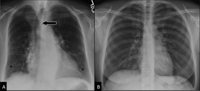
Figure 2. Aging airways.
Comparison between “normal” CXR in an elderly subject (A) and in a young adult (B). Postero-anterior CXR in an 87 years-old woman (A) shows displacement of the trachea (arrow) to the right side, calcifications of the tracheobronchial cartilage, symmetrical bilateral reduction in lung vascularity (more prominent in middle-upper regions), linear and reticular opacities in lung bases (asterisks), and bronchial wall thickening. All these findings cannot be seen in the younger adult (B, 45 years-old woman).

