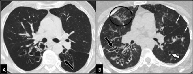
Figure 3. Clinically relevant airway disease.
Comparison between bronchiectases (A) and bronchiolitis due to underlying bronchiectases (b). CT is diagnostic in both cases showing in the first case (A) dilated bronchi with cystic appearance (arrow) whereas in the second case (B) dilated bronchi with cylindrical and varicose (black arrow) appearance, together with bronchial mucoid impaction (black asterisk), thickening of bronchial walls (thin white arrows), centrilobular nodules and consolidations (circle). The patchy and asymmetric distribution of the above-mentioned findings is typical of infectious bronchiolitis. Note the “signet ring sign” (thick white arrow) typical of bronchiectasis, due to the greater diameter of the bronchus in comparison to the adjacent artery.

