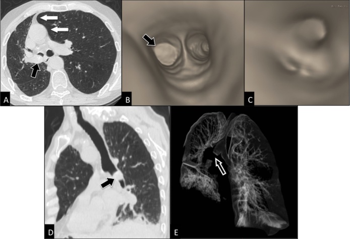
Figure 4. Post-processing imaging techniques: 2D and 3D reconstruction images.
Axial CT scan (A) shows an endobronchial mass (black arrow) within right main bronchus in a 78 years-old man with squamous cell carcinoma, causing partial collapse of right lung with displacement of mediastinum towards right (white arrows). 3D virtual bronchoscopy (B, C) reproduces the bronchoscopic appearance of the mass occluding right main bronchus (arrow in b), but it allows more than bronchoscopy the visualization of the patent bronchi distal to obstruction site (C). Curved 2D MPR (multiplanar reconstructions) image (D) better depicts the cranio-caudal extent of the mass (arrow) into right main bronchus than the axial image (A). CT bronchography (E) allows a global visualization of the aerated lung and shows the site of interruption (arrow) of the tracheobronchial tree.

