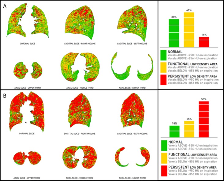
Figure 7. Post-processing imaging techniques: PRM in co-registration analysis.
Co-registration of inspiratory and expiratory CT scans (Imbio LLC, Minnesota, MN) provides a quantitative overview of the lungs (parametric response maps - PRM), with the relative volume of normal parenchyma (green), persistent airway disease (red), and functional airway disease (yellow). This innovative approach allows phenotypization of COPD without the need of an operator, as it is completely automated. Case “A” is a patient with predominant conductive airway disease (47% functional low-density area and 14% persistent low density area), whereas case “B” is a case with predominant emphysema (25% functional low density area and 55% persistent low density area).

