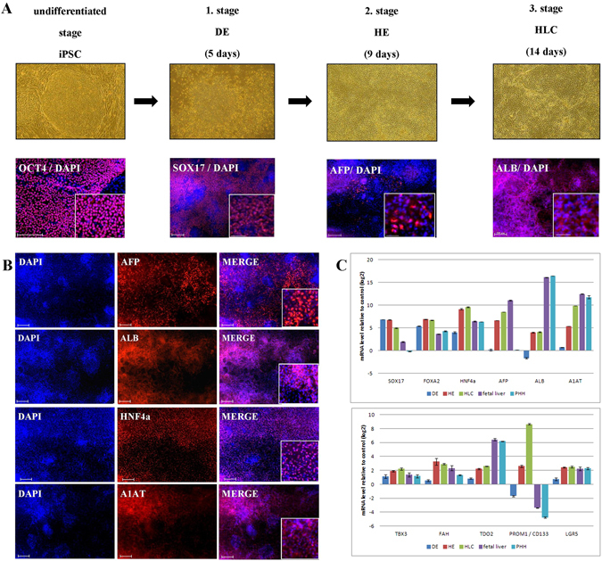
Figure 1.

Derivation of hepatocyte-like cells (HLC) from E-iPSCs. (A) First row phase contrast images of the differentiation stages, from undifferentiated stage the episomal induced pluripotent stem cells (E-iPSCs) to definitive endoderm (DE), then hepatic endoderm (HE) and finally hepatocyte-like cells (HLCs). Second row immunofluorescence-based staining of stage specific proteins overlapped with DAPI (staining of nucleus). Scale bar: 100 μm Alexa Flour 594 (red). (B) Immunofluorescence-based staining of HLC specific proteins AFP, ALB, HNF4α and A1AT. Scale bar: 100 μm Alexa Flour 594 (red). (C) Expression patterns of liver specific marker genes during HLC differentiation compared to fetal liver and primary human hepatocytes (PHH) are shown by quantitative real-time PCR (qPCR). Three biological replicates in technical triplicates of each sample were analyzed. The data were normalized to E-iPSCs. The standard deviation is depicted by the error bars.
