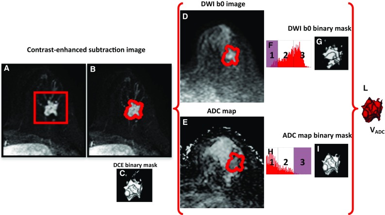
Fig. 1.

Procedure for the semi-automated extraction of the lesion functional volume from MR ADC maps. a The user draws a 3D rectangular box on the lesion on DCE subtraction images; b the enhanced tissue is segmented; c the DCE binary mask of the enhanced tissue is generated; d the DCE binary mask is applied on DWI b0 images; e the DCE binary mask is applied on the ADC maps; f a three compartment k-means algorithm is applied on signal intensity to classify DWI b0 images; g the DWI b0 binary masks is generated by excluding low signal (compartment 1—noise, fat, and fibrous tissue); h a three compartment k-means algorithm is applied on signal intensity to classify ADC maps; i the ADC binary masks is generated by excluding high signal (compartment 3—cyst, necrosis, fluid, normal tissue, and noise); l the probable spatial extension of high cellularity V ADC is the overlap between the DWI b0 and the ADC binary masks
