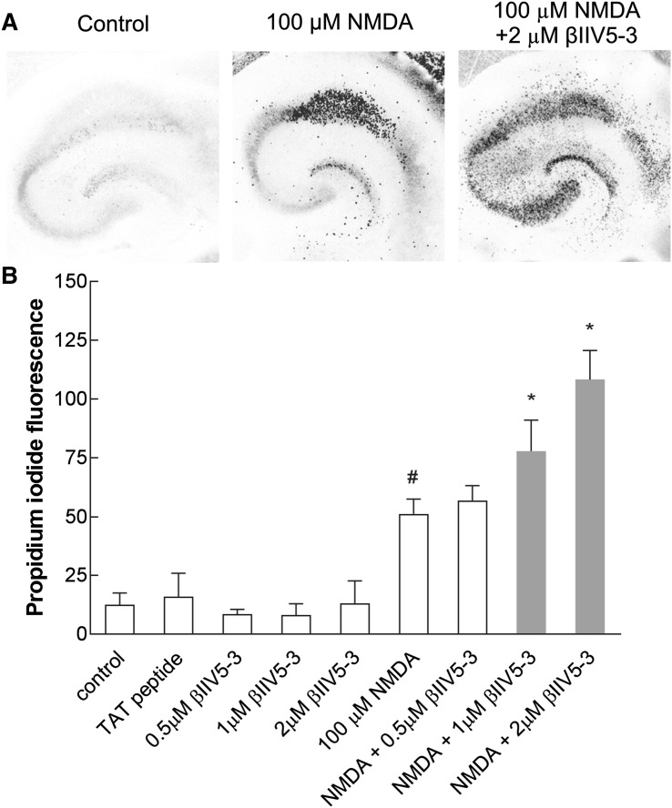
Fig. 4.
βIIV5-3 peptide-enhanced damage initially induced by NMDA in both the CA1 and CA2-4, DG regions of the hippocampus in an in vitro model of ischemia—organotypic hippocampal culture. a Colour-inverted fluorescent images of propidium iodide-stained hippocampal slices 24 h after treatment with 100 μM NMDA alone or together with 2 µM βIIV5-3. b Cell death was measured by reference to propidium iodide fluorescent intensity. The results are expressed as the mean ± SD of cells from three independent experiments. *p < 0.01 versus NMDA, #p < 0.01 versus control

