
Figure 2.
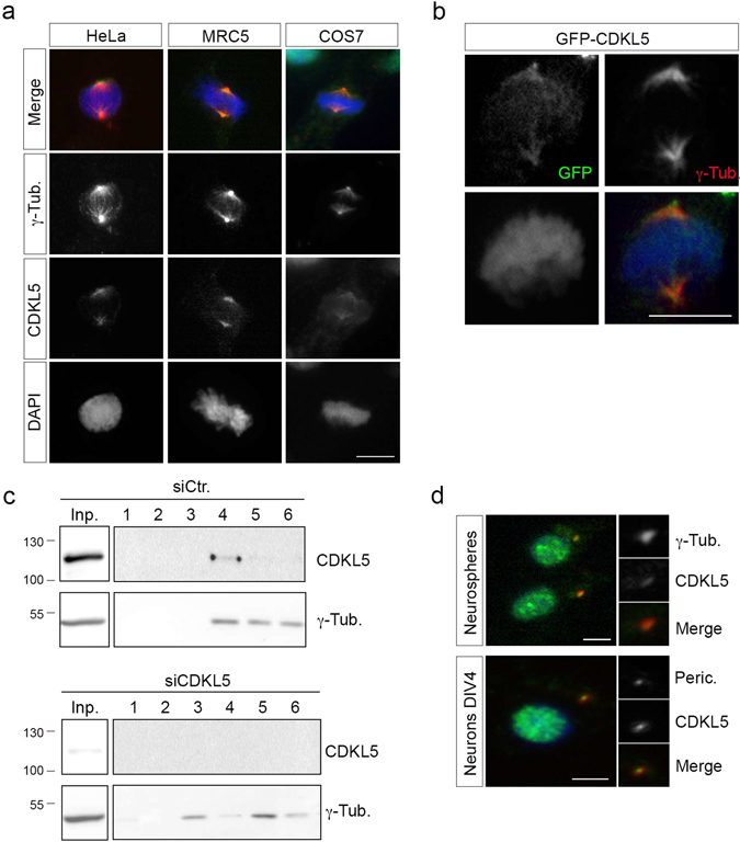
CDKL5 is localized at the centrosome. (a) The indicated cells were stained as in Fig. 1. (b) Exogenously expressed GFP-CDKL5 (green) localizes at the γ-tubulin positive (red) centrosome in HeLa cells. DAPI staining (blue) was used to visualize DNA. (c) HeLa cells were transfected with siCDKL5#1 or a control siRNA (siCtr.); centrosomes were purified 60 h post-transfection and the obtained fractions analyzed by WB with Abs against CDKL5 and γ-tubulin. Input corresponds to approximately 0.6% of the whole cell extract before fractionation. Fractions 1 and 6 are the bottom and top ones, respectively (n = 3 independent experiments). Input and fractions 1–6 are shown as different exposures of the same membrane; full-length blots are presented in Supplementary Figure S7a. (d) Neurospheres (upper) and primary hippocampal neurons (DIV4; lower) were stained with Abs against CDKL5 (polyclonal, green) and either γ-tubulin or pericentrin (both red) and DAPI (blue). The panels to the right show the magnified centrosome. Scale bar, 10 µm.