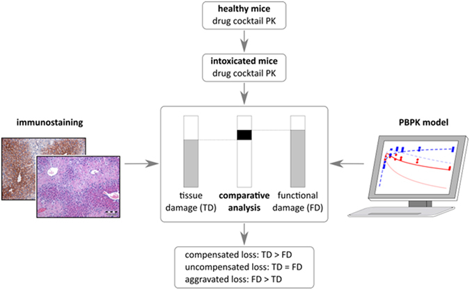
Figure 1.

Overall workflow. In a first step, models for healthy mice were established on the basis of literature knowledge and own experimental data including PK measurements and quantification of enzyme expressing area of the liver lobule. The models were further adjusted to the plasma concentration profiles of intoxicated mice by reducing the clearance capacity of the liver (functional damage, FD). In a complementary approach, the dead cell area of the liver lobule was measured to quantify the tissue damage (TD). The functional damage was then compared to the tissue damage to differentiate between compensated, uncompensated and aggravated loss, respectively, of metabolically active tissue.
