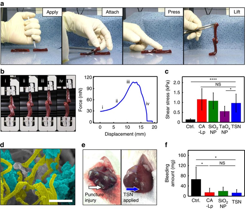
Figure 2. Adhesion strength and hemostatic effect of TSNs in a liver puncture model.
(a) Photographs showing the lap joint preparation procedure for analysing adhesive strength of various glues on biological tissues of two calf liver ribbons: applying the adhesive, attaching by overlapping the ribbons at the region where the adhesive was applied, gently pressing the ribbon for 30 s, and lifting up and moving the joint to be analysed. (b) Photographs showing the lap joint shear test, and a representative force-displacement curve of the lap joint shear test of liver ribbons adhered by TSNs: (i) lifting the lower ribbon, (ii) loading the stress, (iii) ultimate stress point and failure, and (iv) sliding. (c) Adhesive shear stress of lap joint attached without glue (control) or with CA-Lp, SiO2 NPs, TaOx NPs and TSNs. (Data are shown as mean±s.d., n=9, *P<0.05, ****P<0.0001, and NS, not significant P>0.05, one-way ANOVA test.) (d) Scanning electron microscopy (SEM) image showing the adsorbing and entangling interaction between fibrin fibers and TSNs. TSNs and fibrin fiber are highlighted blue and yellow, respectively. Scale bar indicates 500 nm. (e) Photographs of a stab wound after conducting each hemostasis procedure: direct pressure (left), applying TSNs (right). (f) Amount of bleeding during each hemostasis procedure. (Data are shown as mean±s.d., n=5, *P<0.05, NS, not significant P>0.05, one-way ANOVA test.)

