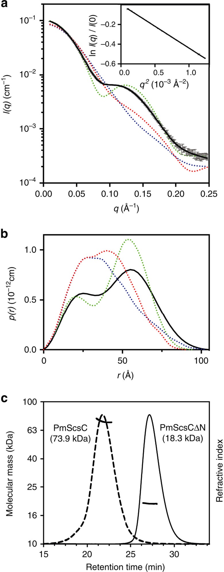
Figure 2. SAXS and MALLS of PmScsC.
(a) Small-angle X-ray scattering data collected from wild type PmScsC (grey) and the calculated scattering profile of the ensemble model overlayed in black (SASBDB: SASDB94). The predicted scattering profile of each of the crystal structures is also shown (dashed lines: PDB: 4XVW compact, red; PDB: 5IDR transitional, blue; PDB: 5ID4 extended, green). The agreement between the experimental data and the ensemble model is excellent, yielding χ2=1.0 (compared to χ2=863.9 (compact); χ2=1222 (transitional); χ2=348.2 (extended)). The Guinier region (inset) of the scattering data is linear, consistent with a monodisperse solution. (b) Pair distance distribution function derived from the scattering data, showing the maximum dimension of the particles in solution is 105 Å. Also shown is the calculated p(r) for each of the crystal structures (dashed lines: compact, red; transitional, blue; extended, green), showing a maximum dimension of 90, 105 and 100 Å, respectively. The p(r) generated from the extended structure (green) is most similar to the experimentally derived p(r), while the other p(r) curves are markedly different. (c) MALLS profile of PmScsC and PmScsCΔN. PmScsC eluted faster (21.5 min) than PmScsCΔN (27.2 min): experimentally determined molecular masses are 74±0.3 kDa for PmScsC (theoretical trimer mass 74.3 kDa) and 18.3±0.3 kDa for PmScsCΔN (theoretical monomer mass 18.1 kDa).

