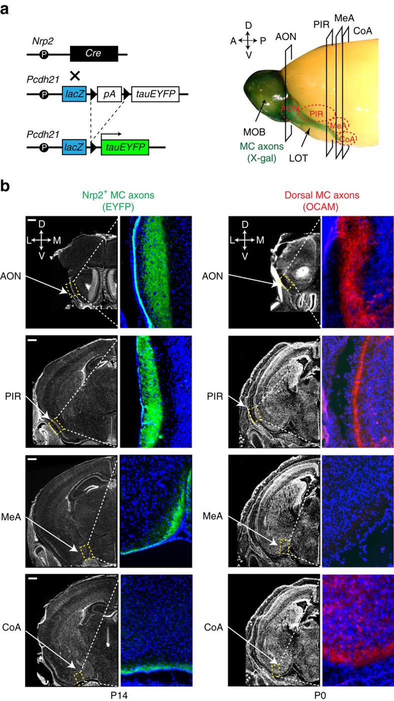
Figure 4. Axonal projection of Nrp2+ MCs to the MeA.
(a) DNA constructs to detect the Nrp2+ MC axons (left). The BAC Tg mouse Nrp2-Cre was crossed with another Tg mouse Pcdh21-lacZ-STOP-tauYFP. Since Nrp2 was difficult to detect in MOB MCs, Tg mice, in which the Nrp2-expressing MCs were labelled with EYFP, were generated. The lacZ was inserted into the construct to detect the promoter activity of Pcdh21 by X-gal staining. Whole mount staining of the MOB is shown (right). Locations of coronal sections (AON, PIR and MeA) analysed in b are indicated. (b) Detection of Nrp2+ MC axons in the OC. Coronal sections of the OC at P14 were immunostained with anti-GFP antibodies to detect EYFP expressed in the Nrp2+ MCs (left). Nrp2+ ventral MC axons (green) project to the AON, PIR, MeA and CoA. Coronal OC sections at P0 were immunostained with anti-OCAM antibodies (right). OCAM+ dorsal MC axons (red) project to the AON and PIR, but not to the MeA. n=6. Scale bar, 100 μm. Nuclei are counterstained with DAPI in blue.

