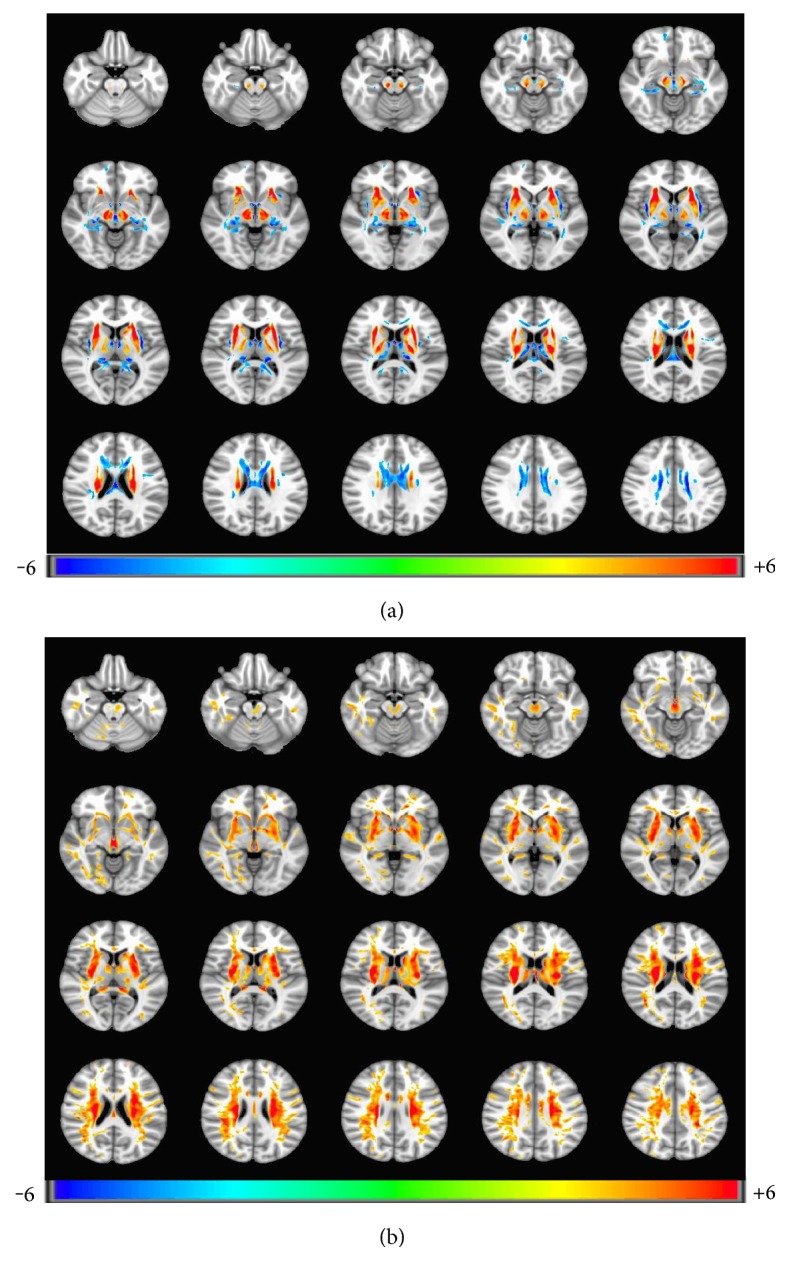
Figure 1.

(a) FA parameter differences of brain regions between patients and controls (FDR simulation, (p = 0.001, α = 0.05, cluster size = 326)). Patients showed increased FA in the bilateral head of the caudate nucleus, lenticular nucleus, ventral thalamus, brain stem, and decreased in mediodorsal thalamus and bilateral extensive matter. (b) MD parameter differences of brain regions between patients and controls (FDR simulation, (p = 0.001, α = 0.05, cluster size = 326)). Patients showed increased MD in the bilateral head of the caudate nucleus, lenticular nucleus, claustrum, thalamus, brain stem, and extensive white matter.
