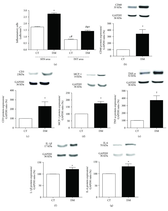
Figure 3.
(a) Total inflammatory cell count in SEN and INT areas. HE-stained sections, under 400x magnification. θp < 0.05 versus the DM SEN area; ∗p < 0.01 versus the CT SEN area, #p < 0.01 versus the DM SEN area; †p < 0.01 versus the CT INT area. Data expressed in mean ± SEM. Western blot protein expression analysis depicting CD68 (b), CD3 (c), MCP-1 (d), TNF-α (e), IL1-β (f), and IL-6 (g) was performed 8 weeks after diabetes induction. ∗p < 0.05 and †p < 0.001 versus the CT group. Data expressed in mean ± SEM in all plots.

