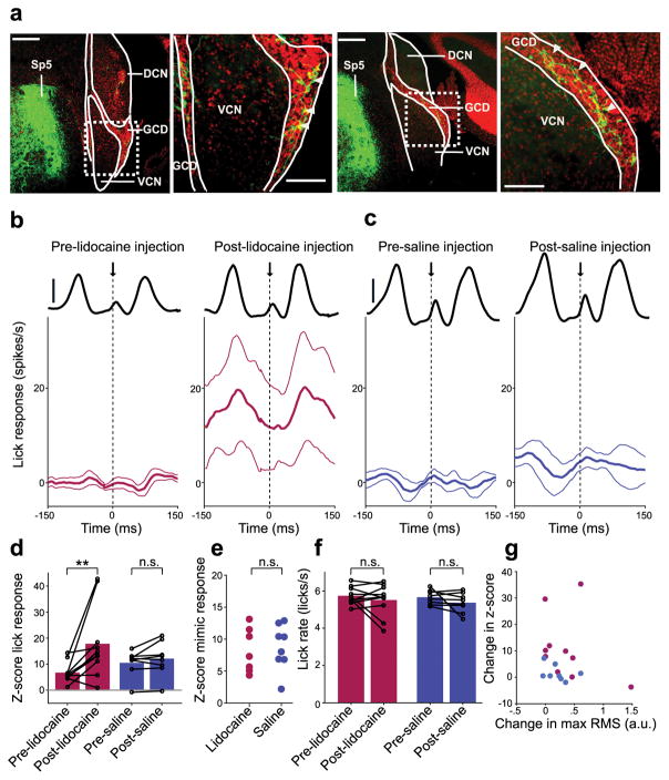
Figure 5. A role for the spinal trigeminal nucleus in cancelling self-generated sounds in DCN.
(a) Labeled mossy fibers were observed in DCN granule cell domains (GCD) after injection of an anterograde viral tracer (AAV2-GFP) into the ipsilateral Sp5. Scale bars: 200 μm. Right, higher magnification views of areas indicated by dotted rectangle. Scale bars: 100 μm. White arrowheads indicate labeled mossy fibers in GCDs. (b,c) Lick-triggered response of DCN cells before (left) and after (right) injection of lidocaine (b, n = 10) or saline (c, n = 8) into Sp5. Thin lines are s.e.m. Solid black lines show the RMS amplitude of the licking sound (scale bars: 1 a.u.). (d) Lidocaine injection resulted in a significant increase in z-scored lick responses in DCN units (P = 0.0098, Wilcoxon Signed Rank Test, red) while no significant increases in z-scored lick responses occurred after saline injection (P = 0.31, Wilcoxon Signed Rank Test, purple). (e) Auditory responses to the mimic were not significantly different in lidocaine and saline groups (P = 0.87, Wilcoxon Rank Sum Test). (f) Lick rate did not differ before and after injection of lidocaine (P = 0.77, Wilcoxon Signed Rank Test) or saline (P = 0.25, Wilcoxon Signed Rank Test). (g) Changes in z-score lick responses were not correlated with changes in the maximum RMS of the licking sound after lidocaine injection (red, P = 0.36, linear regression t-test). Changes in the maximum RMS of the licking sound did not differ between lidocaine and saline groups (P = 0.51, Wilcoxon Rank Sum Test).

