Fig. 4.

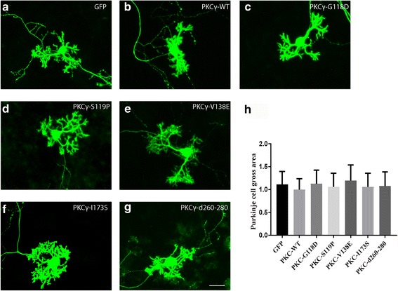
Morphology of Purkinje cells transfected with PKCγ C1 or C2 domain mutants. The morphology of Purkinje cells was analyzed after 2 weeks in culture. a Purkinje cells transfected with GFP control vector. b Purkinje cells transfected with PKCγ WT. c Purkinje cells transfected with PKCγ-G118D, d PKCγ-S119P, e PKCγ-V138E or f PKCγ-I173S. g Purkinje cells transfected with PKCγ Δ260–280. Scale bar in G = 50 μm. h Quantification of the Purkinje cell area. The dendritic tree size of Purkinje cells transfected with C1 or C2 domain mutations was not significantly different from control Purkinje cell transfected with the empty vector expressing GFP only or from PKCγ WT transfected Purkinje cells. Data are shown as the mean ± S.D. of at least 40 Purkinje cells. The value for Purkinje cells transfected with PKCγ WT was set as 100%
