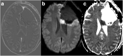
Fig. 1.

a shows a postoperative subtraction, b a postoperative diffusion-weighted image (DWI, b 1000), and c the corresponding apparent diffusion coefficient (ADC) map. Images a–c show an example of a postoperative ischemia with restricted diffusion in the genu of the corpus callosum in a patient diagnosed with an anaplastic oligodendroglioma
