
Fig. 1.
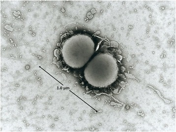
Photomicrograph of N. lactamica Y92–1009. This image was obtained with transmission electron micrography and displays the diplococcoid nature of the N. lactamica Y92–1009 cell. The size of the cell is indicated in micrometres (μm)
Official websites use .gov
A
.gov website belongs to an official
government organization in the United States.
Secure .gov websites use HTTPS
A lock (
) or https:// means you've safely
connected to the .gov website. Share sensitive
information only on official, secure websites.
Photomicrograph of N. lactamica Y92–1009. This image was obtained with transmission electron micrography and displays the diplococcoid nature of the N. lactamica Y92–1009 cell. The size of the cell is indicated in micrometres (μm)