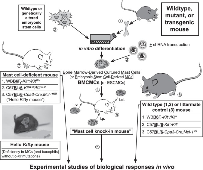
Fig. 1.
Making “mast cell knock-in mice.” (1) Mast cells can be generated from bone marrow cells (or other hematopoietic cells, e.g., those in the fetal liver) from wild-type mice or from mutant or transgenic mice with specific genetic alterations of interest (50–52). Alternatively, (2) embryonic stem (ES) cell–derived cultured mast cells (ESCMCs) can be generated from wild-type or genetically altered ES cells (53,54), or (3) various mast cell populations can be transduced in vitro with shRNA to diminish expression of specific genes of interest (15,55). (4) Such bone marrow–, ES cell–, (or fetal liver–) derived cultured mast cells, or shRNA-transduced mast cells, can then be transplanted into mast cell–deficient c-kit mutant mice, such as WBB6F1-KitW/KitW-v mice (52,56) or C57BL/ 6-KitW-sh/KitW-sh mice (57,58), or into C57BL/6-Cpa3-Cre;Mcl-1fl/fl mice (59) (which we informally refer to as “Hello Kitty mice”, which have wild-type c-kit), to produce mast cell knock-in mice. Note: Mouse bone marrow–derived cultured mast cells (BMCMCs) can be injected into genetically mast cell-deficient mice intravenously (i.v.), intraperitoneally (i.p.), or intradermally (i.d.), or into the joints or meninges, etc., but there is a more limited experience with the engraftment of other types of MCs, such as EMCMCs, than with BMCMCs. (5) A suitable interval is then allowed for engraftment and phenotypic “maturation” of the adoptively transferred mast cells (the length of this interval can be varied based on the route of mast cell transfer, the anatomical site of interest, the particular biological response being analyzed, etc.). The importance of mast cell function(s) in biological responses can be analyzed by comparison of the responses in the appropriate wild-type or littermate control mice (6), the corresponding mutant mast cell-deficient mice (7), and selectively mast cell–engrafted mutant mice (mast cell knock-in mice) (8). The contributions of specific mast cell products (surface structures, signaling molecules, secreted products, and so on) to such biological responses can be analyzed by comparing the features of the responses of interest in mast cell knock-in mice engrafted with wild-type mast cells versus mast cells derived from mice or ES cells that lack or express genetically altered forms of such products or that have been transduced with shRNA to silence the specific genes that encode these products. An important part of the analysis of the mast cell knock-in mice used in particular experiments is to assess the numbers and anatomic distribution, and, for certain experiments, aspects of the phenotype, of the adoptively-transferred mast cells, as, depending on the type of in vitro–derived mast cells used, the route of administration, and other factors, these may differ from those of the corresponding native populations of mast cells in the corresponding wild type mice (14,60,61). [This is a modified version of Figure 2 from Metz M, Grimbaldeston MA, Nakae S, et al. Mast cells in the promotion and limitation of chronic inflammation. Immunol Rev 2007;217:304-28 (ref. 60), reprinted with the permission of the publisher, John Wiley and Sons.]

