Figure 3.
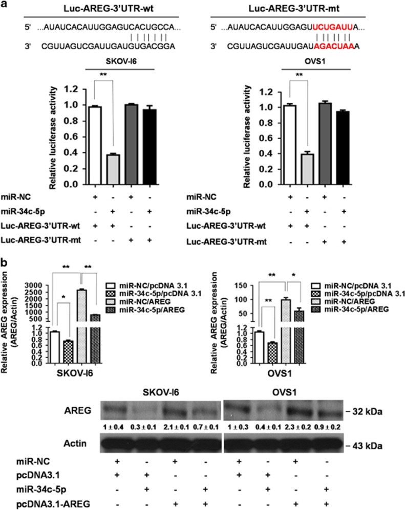
Identification of AREG as the direct target of miR-34c-5p. (a) Effect of miR-34c-5p on Luc-AREG-3′UTR-wt (wild type) and Luc-AREG-3′UTR-mt (mutant) luciferase reporters in SKOV-I6 and OVS1 cells. Top, the Luc-AREG-3′UTR-wt sequence and the Luc-AREG-3′UTR-mt sequence in which the sequence in red was mutagenized to abolish the binding between miR-34c-5p and AREG-3′UTR. Bottom, Luciferase reporter assay showed decreased activity of more than 50% after cotransfection with miR-34c-5p and Luc-AREG-3′UTR-wt in SKOV-I6 and OVS1 cells. The activity of Luc-AREG-3′UTR-mt was not significantly affected by miR-34c-5p. Histograms represent means±s.d. from three independent experiments (*P<0.05, **P<0.01). (b) miR-34c-5p inhibited AREG in both mRNA and protein expression levels in SKOV-I6 and OVS1 cells. Top, the mRNA expression levels of AREG in both cell lines were measured by qRT–PCR from cells transfected with the indicated plasmids. Histograms represent means±s.d. from three independent experiments (*P<0.05, **P<0.01). Bottom, the protein expression levels as reflected by western blotting of AREG in both cell lines transfected with the indicated plasmids are shown. β-actin was used as an internal control. Relative band intensity was quantified by ImageJ 1.42 and was represented with normalized mean±s.e. (N=3) below each band.

