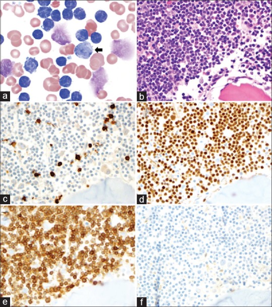
Figure 1.

(a) Peripheral smear (×1000) demonstrating a predominance of small, mature lymphocytes, scattered smudge cells, and a rare large cell with morphology consistent with a prolymphocyte (arrow); (b) Bone marrow biopsy (×400) demonstrating sheets of small mature lymphocytes; (c) CD3 stains a few scattered, background T-cells (×400); (d) PAX-5 stains the majority of small lymphocytes (×400); (e) PAX-5-positive B-cells co-express CD5 (×400); (f) B-cells are negative for BCL-1 (×400)
