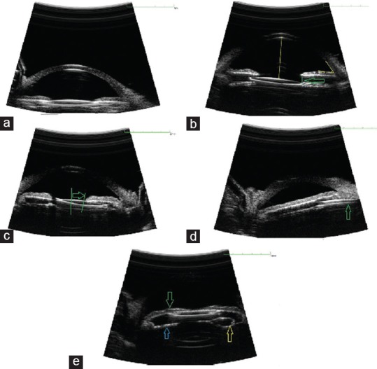
Figure 1.

Ultrasound biomicroscopy sample images of a single-piece intraocular lens (IOL) implanted into the ciliary sulcus. (a) Well-positioned posterior chamber (PC)-IOL in the ciliary sulcus, (b) tilt of IOL's optic (displacement of one edge of IOL's optic from margin of pupil >100 µm [arrow]), (c) IOL decentration (displacement of the center of IOL's optic from the center of the pupil >0.5 mm [distance between 2 green lines]), (d) iris root flanking by the IOL haptic (arrow), (e) anterior bowing of iris (green arrow) and Soemmering ring formation (yellow and blue arrows).
