Figure 3.

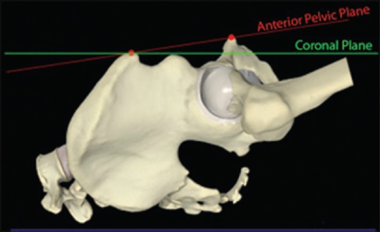
Schematic representation on saw bones demonstrating pelvic tilt - the difference between the anterior pelvic plane and the coronal plane

Schematic representation on saw bones demonstrating pelvic tilt - the difference between the anterior pelvic plane and the coronal plane