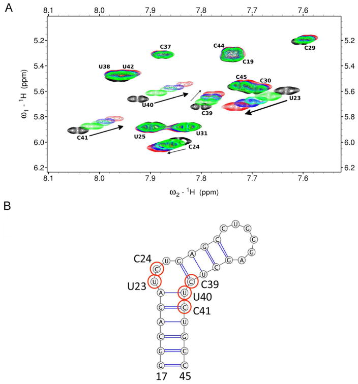
Figure 3.
Overlaid 2D TOCSY NMR spectra of pyrimidine residues of TAR (A) with increasing concentrations of 2. Indicated are free TAR (black) and TAR:2 ratio of 1:1 (green), 1:2 (light blue), 1:3 (dark blue), and 1:5 (red). Arrows indicate the direction of chemical shift changes upon addition of 2. (B) Secondary structure of 29nt TAR hairpin with bases perturbed in the presence of 2 highlighted with red circles.

