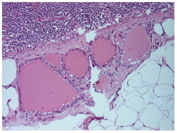
Fig. 3.

A collection of < 10, colloid containing, thyroid follicles lined by cuboidal epithelium in the capsule of a level IIA, cervical lymph node from a ND for pT4a N1, SCC of the left mandibular alveolus in a male aged 56 years. Lymphoid parenchyma and perinodal fat are seen at the upper left and lower right of the picture, respectively.
