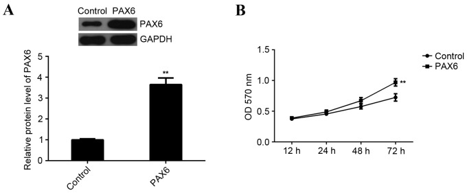
Figure 6.
PAX6 is involved in miR-375-mediated proliferation of MCF-7 cells. (A) Western blot analysis was performed to examine the expression of PAX6 protein in MCF-7 cells transfected with PAX6 plasmid. GAPDH was used as an internal reference. (B) MTT assay to determine the rate of cell proliferation in each group. **P<0.01 vs. Control. PAX6, paired box 6; miR, microRNA; MCF, Michigan Cancer Foundation; GAPDH, glyceraldehyde 3-phosphate dehydrogenase; OD, optical density; Control, non-transfected MCF-7 cells.

