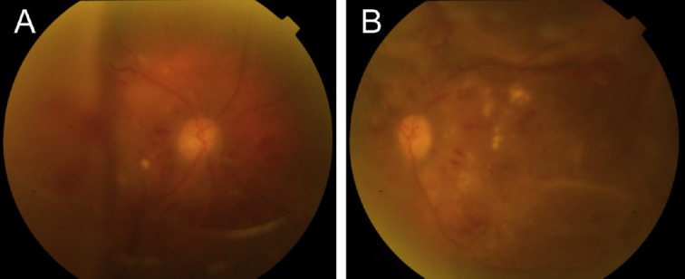
Figure 2.

Color fundus photos at initial presentation showed bullous retinal detachments in (A) the right eye and (B) the left eye. Diffuse retinal arteriolar narrowing, increased vascular tortuosity, multiple dot and blot retinal hemorrhages, and macular exudations could also be found in both eyes.
