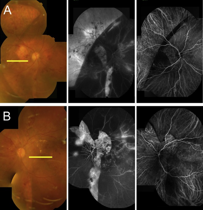
Figure 4.
Comparison of color fundus photos (left), fluorescein angiographies (middle), and indocyanine green angiographies (right) 2 weeks after initial presentation. (A) A large RPE defect can be found at the temporal quadrants of the right eye. Vertical RPE defects can be found at the temporal and inferior sides of the optic disc. (B) RPE defects can be found at the superior and temporal sides of the optic disc. The RPE defects correspond to the sharply defined hyperfluorescent areas on the fluorescein angiography and the increased visibility of choroidal vessels on the indocyanine green angiography. RPE = retinal pigment epithelial.

