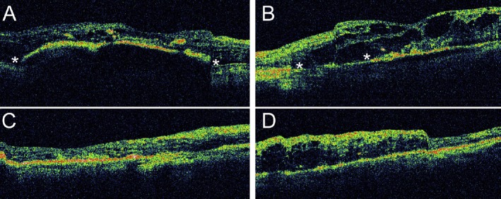
Figure 5.
OCT of (A) the right eye and (B) the left eye 2 weeks after initial presentation. The corresponding locations of the scans are marked in the color fundus photos in Figure 4. Asterisks (*) indicate the locations of the RPE defects. Intraretinal fluid and exudates can be found in both eyes. OCT of (C) the right eye and (D) the left eye 3 months after initial presentation. The macular edema in the right eye improved after an intravitreal injection of bevacizumab. The left eye received no treatment, and its macular edema persisted. OCT = optical coherence tomography; RPE = retinal pigment epithelial.

