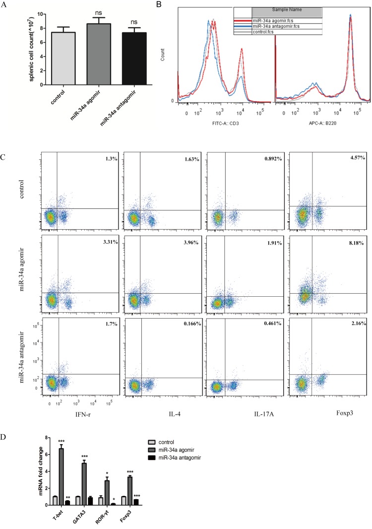
Figure 4.
Effect of modification of miR-34a function on peripheral lymphocyte distribution. (A) Splenocytes were counted, and no significant changes were seen in the spenic cell count. (B) Histogram showing the percentages of CD3+ and B220+ cells in the splenocytes. (C) Flow cytometry of Th subsets. (D) mRNA expression of transcriptional factors in the lymph nodes. *P<0.05, ns means no significence, **P<0.01, ***P<0.001 vs. control group.

