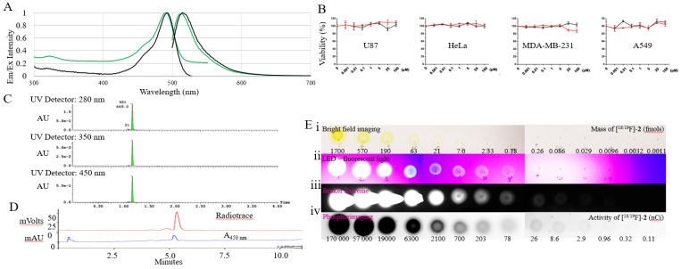
Figure 1.
Spectral and radioactive properties of [18/19F]-2. (A) Excitation and emission spectra of 5-6 μM solutions of fluorescein (black) and [18/19F]-2 (green) in 1x PBS, pH 7.4 measured on a Cary Eclipse spectrophotometer, with 5 nm slit widths, and excitation at 490 nm. (B) In vitro cell viability tests performed with compound 2 (red line) and fluorescein (control) 24 hour on U87, Hela, MDA-MB-231, and A549 immortal cell lines using a CellTiter 96® AQueous One Solution Cell Proliferation Assay (MTS, G3582, Promega). Toxicity is not observed. (C) UV-Vis (280, 350, and 450 nm) UPLC-MS of 2 on a waters Acuity UPLC and a Phenomenex Luna Kinetex 1.7 µm EVO C18 50 x 2.1 mm column (00B-4726-AN), with a 1.5 min, a10-90% H2O:ACN (0.05% TFA) elution gradient indicating a pure synthesis of 2. (D) Reverse phase HPLC of radiolabeled [18F]-2 on a Varian HPLC, using an Waters SunfireTM C18 3.5 μm 4.6 x 50 mm column (186002551), a10-90% H2O:ACN (0.05% TFA) elution gradient and a flow rate of 2 mL/min. (E) 10 µL 3x dilution series of [18/19F]-2 beginning at 70 µM (1.7 pmols) and 170 µCi of [18/19F]-2 (first spot). A 10 µl volume was plated onto glass-backed TLC plate model. Imaging was performed using (i) bright-field; (ii) a UV-sight hand-held 4.5 volt LED black-light (fluorescence); (iii) a Bruker Xtreme optical imaging device (fluorescence: 1 sec acquisition using excitation and emission filters set at λex= 450 nm and λem= 535 nm); (iv) and a phosphorimager.

