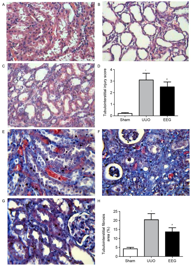
Figure 2.
Morphological analysis of renal histology. Hematoxylin-eosin staining was performed to evaluate tubulointerstitial injury (A-D) and Masson staining was performed to evaluate tubulointerstitial fibrosis (E-H). (A and E) No marked histological abnormalities were observed in the sham group. (B and F) Renal fibrosis was clearly visible in the UUO group. (C and G) EEG markedly decreased tubulointerstitial injury and inflammatory cells infiltration. (D) Statistical analyses of tubulointerstitial injury scores. (H) Statistical analyses of relative percentages of tubulointerstitial fibrosis. Images were captured at a magnification of ×400. Data are presented as the mean ± standard deviation (n=10). *P<0.05 vs. sham group; #P<0.05 vs. UUO group. UUO, unilateral ureteral obstruction; EEG, ethanol extract of gardenia fruits.

