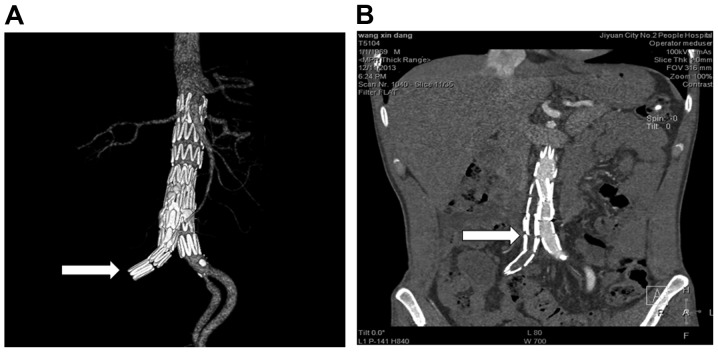
Figure 2.
Three-dimensional reconstruction of 64-slice computer tomographic angiography images of the 43-year-old patient (case no. 6) at 1 year after operation showing (A) the artery and (B) the full body. Occlusion in the right iliac artery was observed (arrows). A low-density shadow was visible, indicating the formation of thrombosis.

