
Figure 2.
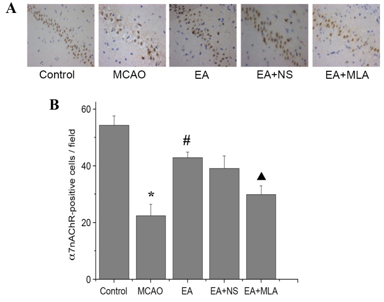
EA increases the expression of α7nAChR in the hippocampus. Distribution of α7nAChR-positive cells in the CA1 region of the hippocampus in the five groups stained with DAB (A) observed under a microscope (magnification, ×400) and (B) quantified using an image analysis system. *P<0.01 vs. control, #P<0.01 vs. MCAO and ▲P<0.01 vs. EA + NS. Data are expressed as mean ± standard error of the mean (n=5). EA, electroacupuncture; α7nAChR, α7 nicotinic acetylcholine receptor; MCAO, middle cerebral artery occlusion; NS, normal saline; MLA, methyllycaconitine; DAB, 3,3′-Diaminobenzidine.