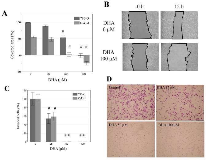
Figure 3.
DHA suppresses the migration and invasion of renal cancer cells. (A) Wound healing data obtained from disruption of Caki-1 and 786-O cell monolayers with a pipette tip following treatment with 0, 25, 50 and 100 µM DHA for 12 h. (B) Representative photomicrographs showing wound closure in Caki-1 cells. Magnification ×200. (C) Data obtained from a Matrigel invasion assay of Caki-1 and 786-O cells after incubation with DHA (0, 25, 50 and 100 µM) for 24 h. (D) Representative photomicrographs indicating Caki-1 cell invasion, magnification ×100. #P<0.001 vs. untreated control cells of respective cell lines. DHA, docosahexaenoic acid; Caki-1 and 786-O, renal cancer cell lines.

