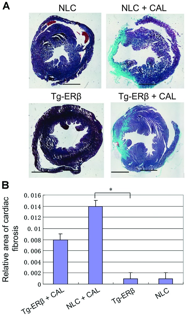
Figure 3.
Masson staining of paraffin sections was performed on NLC, NLC + CAL, Tg-ERβ, Tg-ERβ + CAL, respectively (myocardial interstitium ×100 magnification, perivascular ×200 magnification). Image-Pro6.0 software was used to analyze the cardiac fibrosis area quantitatively and statistical results were obtained. Compared with the NLC + CAL group, the area of coronary artery and myocardial interstitial fibrosis was significantly reduced in the Tg-ERβ + CAL group, and the difference was statistically significant, *P<0.05. ERβ, estrogen receptor β; NLC, non-transgenic littermate control; CAL, coronary artery ligation.

