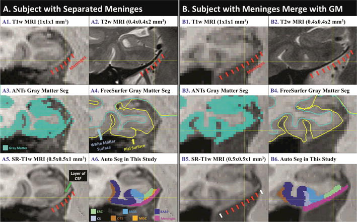
Fig. 1.

Coronal slices of T1w MRI (A1, B1) and T2w MRI (A2, B2) from the same subjects with meninges highlighted by red arrows, which are commonly segmented as gray matter by the state-of-the-art algorithms, including ANTs (A3, B3) and FreeSurfer (A4, B4). Super-resolution technique increases visibility of this structure (A5, B5) that helps automatic segmentation (A6, B6). A layer of CSF between meninges and GM is visible in SR-T1w MRI in some cases (A5) but not in all (B5).
