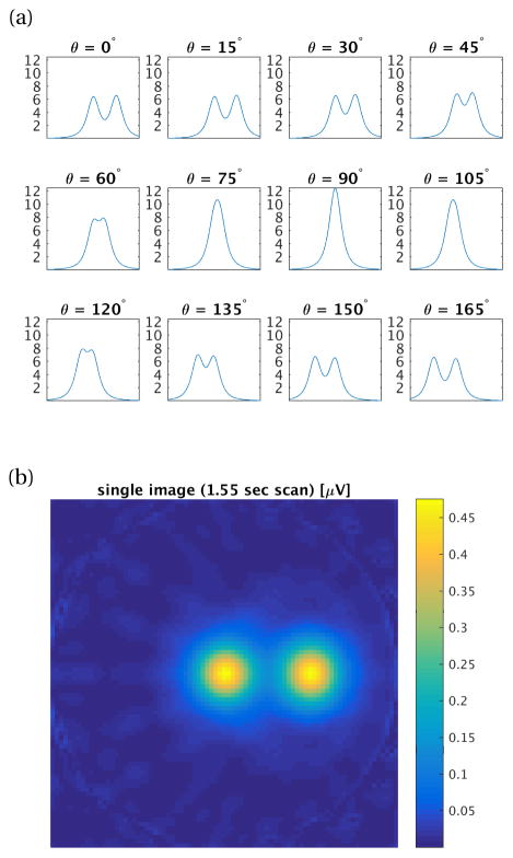
Figure 2.
(a) Simulated projections [μV] for 12 angles from 0° to 180° about two 50 μg Fe samples in 2.3 mm3 volumes located at (x, y, z) = (0,0,0) and (5 cm,0,0). Drive field: 25 mT at 10 kHz, gradient FFL: 1.5 T/m. Projection axis: [−10 cm, 10 cm], 81 points. Signal received with human-head size solenoid receive coil with 25 turns (coil (c) in Tab. 1), signal filtered to 3rd harmonic. Added noise corresponds to Nyquist noise in receive coil at sampling BW100 kHz (9.77 nV). 1.6ms scan per point, 1.55 sec imaging time. (b) Reconstructed axial slice image using the 12 projections from (a).

