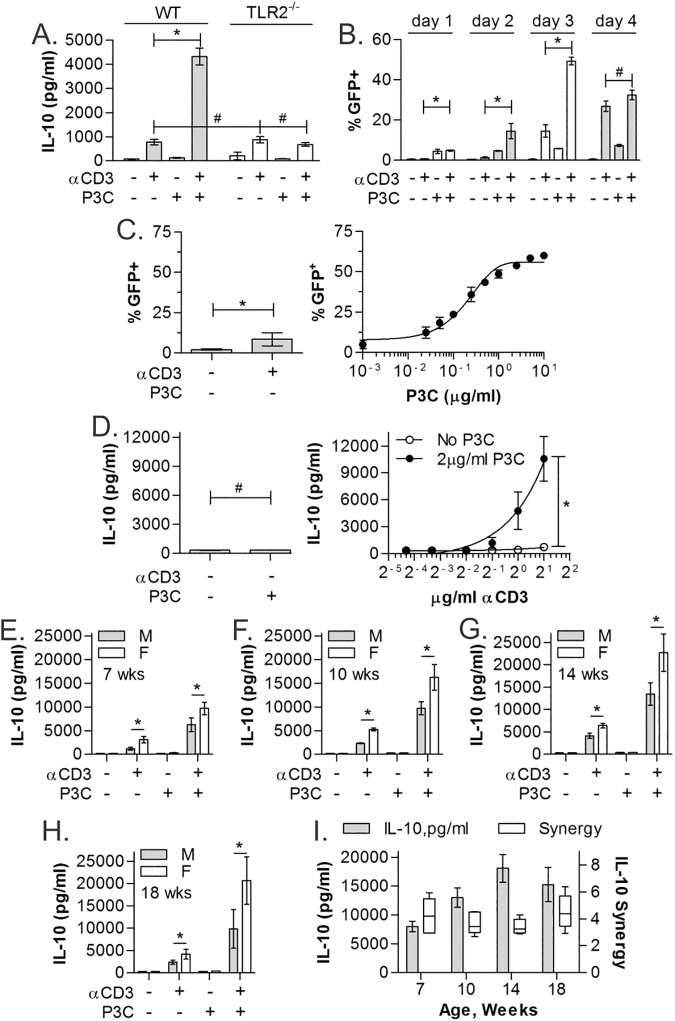
Fig 2. TLR2-induced IL-10 is dose-dependent and independent of age and sex.
(A) CD4+ T cells from WT (gray) and TLR2-/- (white) mice isolated using MACS were stimulated as indicated for 3 days and IL-10 quantified by ELISA showing ablation of IL-10 synergy without TLR2. (B) IL-10-GFP reporter CD4+ T cells isolated using MACS were used to test kinetics of the IL-10 response over 4 days. Peak synergy was seen on day 3. (C) IL-10-GFP reporter CD4+ T cells isolated using MACS were stimulated with αCD3 (2μg/ml) and varying concentrations of P3C for 3 days and analyzed by flow cytometry, revealing strong dose-dependence on P3C. (D) WT CD4+ T cells isolated using MACS were stimulated with P3C (2μg/ml) and varying concentrations of αCD3 for 3 days and analyzed by ELISA, also showing strong dose-dependence on αCD3. (E-H) WT CD4+ T cells were isolated from males and females of varying ages ranging from 7 weeks to 18 weeks using MACS and stimulated as before for 3 days. (I) Overall IL-10 amplitude increased through 14 weeks of age, and female mice produced more IL-10 than their male counterparts, however, the degree of IL-10 synergy (fold increase in IL-10 with αCD+P3C over the sum of the individual stimulations) was unchanged across age and gender. All data are n≥3, unless otherwise noted; *p<0.05; #p>0.05.

