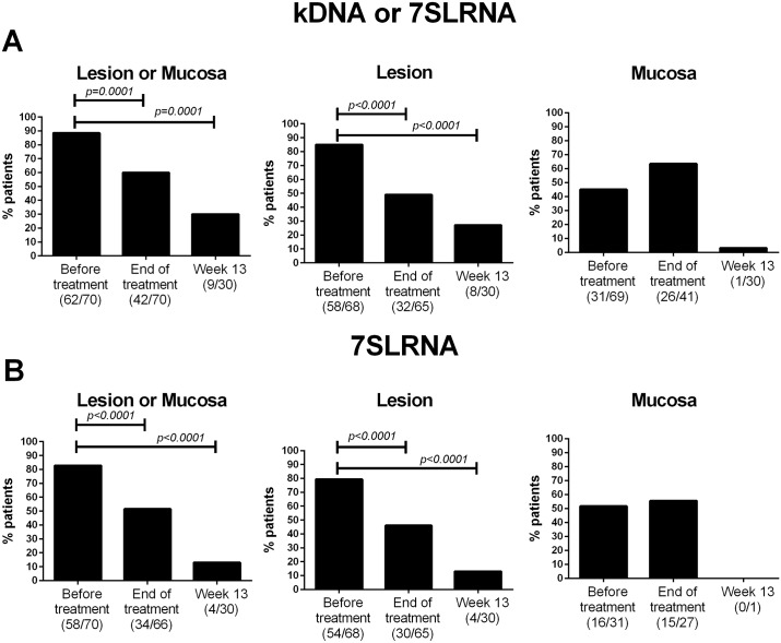
Fig 2. Frequency of detection of Leishmania in samples from CL patients before and after treatment.
Presence of Leishmania was determined by detection of Leishmania kDNA or 7SLRNA in lesion and mucosal samples (A) and parasite viability (B) was determined by detection of 7SLRNA transcripts. Graphs represent frequency of positivity in at least one sample (left panels), or independently in lesions (center panels) and in any mucosal tissue (right panels). Data are shown as relative frequencies based on the total number of patients at each sampling time. Differences were analyzed by the McNemar’s test.

